Amyloid Angiopathy Mri Sumer S Radiology Blog

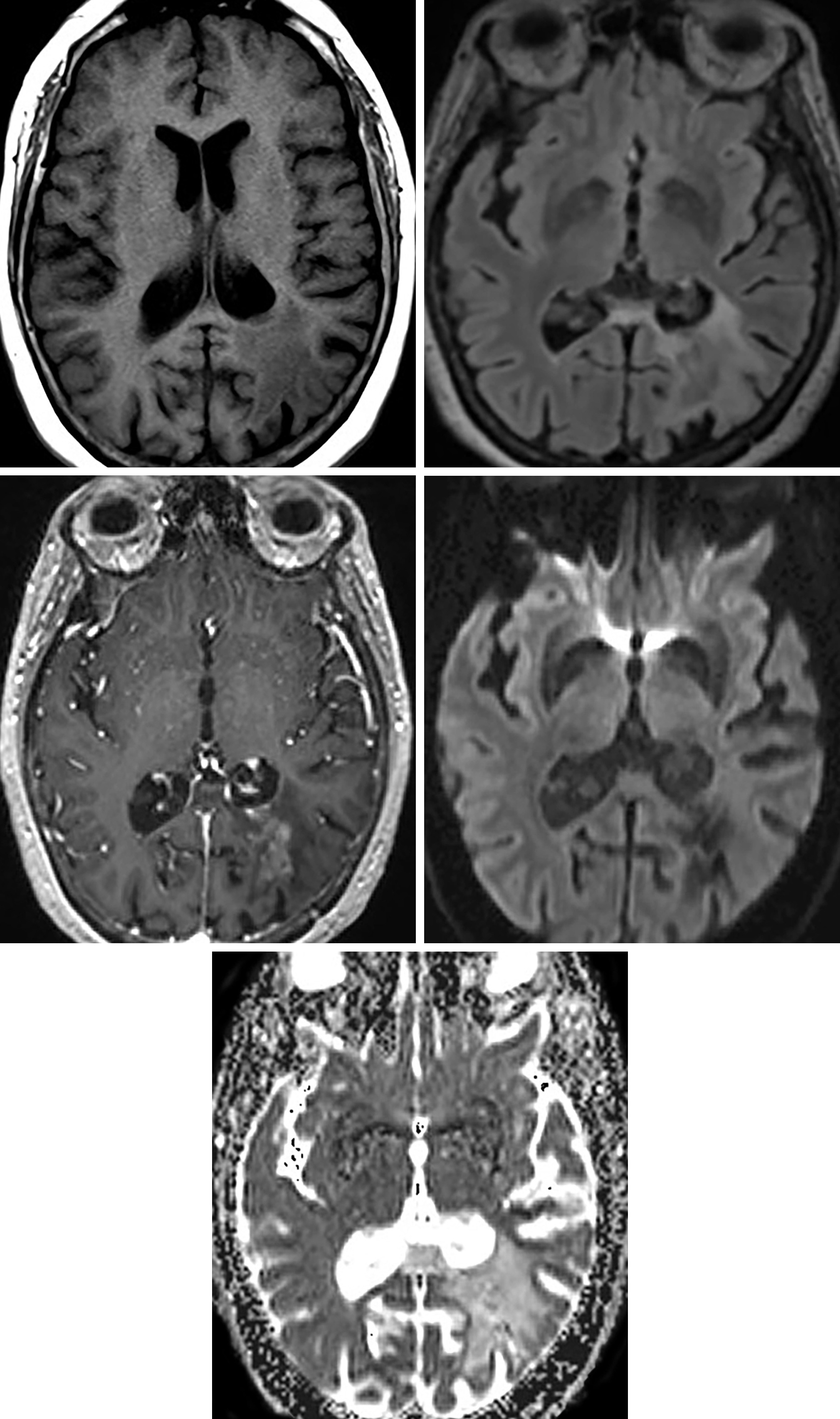
Amyloid Angiopathy Mri Sumer S Radiology Blog Friday, september 06, 2013 amyloid angiopathy , neuroradiology. 64 year old male with a known chronic kidney dysfunction & myocardial ischaemia presence with altered sensorium. the mri brain shows extensive parenchymal bleeds of varying sizes in the entire cerebral, cerebellar parenchyma, basal ganglia & brain stem with t1 hyperintensity and. Cerebral amyloid angiopathy is a frequent incidental finding, found on screening gradient recalled echo imaging in up to 16% of asymptomatic elderly patients 4. autopsy studies have found a prevalence of approximately 5 9% in patients between 60 and 69 years, and 43 58% in patients over the age of 90 years 4 .

Amyloid Angiopathy Mri Sumer S Radiology Blog The diagnostic criteria for possible or probable inflammatory cerebral amyloid angiopathy require age ≥40 years 4. in general, the same patient group affected by cerebral amyloid angiopathy is affected, and thus most patients are elderly, typically 60 80 years of age. however, the average patient is a little younger than in non inflammatory. Cerebral amyloid angiopathy (caa) is an important but underrecognized cause of cerebrovascular disorders that predominantly affect elderly patients. caa results from deposition of β amyloid protein in cortical, subcortical, and leptomeningeal vessels. this deposition is responsible for the wide spectrum of clinical symptoms and neuroimaging findings. many cases of caa are asymptomatic. The boston criteria 2.0 were proposed in 2022 in order to better include leptomeningeal and white matter characteristics into the diagnoses of probable and possible cerebral amyloid angiopathy (caa) 1. they consist of combined clinical, imaging and pathological parameters, and are based upon the original boston criteria and modified boston. Cerebral amyloid angiopathy (caa) refers to the deposition of b amyloid in the media and adventitia of small and mid sized arteries (and less frequently, veins) of the cerebral cortex and the leptomeninges. it is a component of any disorder in which amyloid is deposited in the brain, and it is not associated with systemic amyloidosis.

Amyloid Angiopathy Mri Sumer S Radiology Blog The boston criteria 2.0 were proposed in 2022 in order to better include leptomeningeal and white matter characteristics into the diagnoses of probable and possible cerebral amyloid angiopathy (caa) 1. they consist of combined clinical, imaging and pathological parameters, and are based upon the original boston criteria and modified boston. Cerebral amyloid angiopathy (caa) refers to the deposition of b amyloid in the media and adventitia of small and mid sized arteries (and less frequently, veins) of the cerebral cortex and the leptomeninges. it is a component of any disorder in which amyloid is deposited in the brain, and it is not associated with systemic amyloidosis. The history of how to diagnosis cerebral amyloid angiopathy (caa) tells the story of the disease itself. caa is defined by histopathology—deposition of β amyloid in the cerebrovasculature—and through the 1980s the disorder was only diagnosed in patients with available brain tissue from hematoma evacuation, biopsy, or most commonly postmortem examination. 1 introduction of the imaging. Abstract. cerebral amyloid angiopathy (caa) is a cerebral small vessel disease caused by β amyloid (aβ) deposition at the leptomeningeal vessel walls. it is a common cause of spontaneous intracerebral hemorrhage and a frequent comorbidity in alzheimer’s disease. the high recurrent hemorrhage rate in caa makes it very important to recognize.

Amyloid Angiopathy Mri Brain The history of how to diagnosis cerebral amyloid angiopathy (caa) tells the story of the disease itself. caa is defined by histopathology—deposition of β amyloid in the cerebrovasculature—and through the 1980s the disorder was only diagnosed in patients with available brain tissue from hematoma evacuation, biopsy, or most commonly postmortem examination. 1 introduction of the imaging. Abstract. cerebral amyloid angiopathy (caa) is a cerebral small vessel disease caused by β amyloid (aβ) deposition at the leptomeningeal vessel walls. it is a common cause of spontaneous intracerebral hemorrhage and a frequent comorbidity in alzheimer’s disease. the high recurrent hemorrhage rate in caa makes it very important to recognize.

Amyloid Angiopathy Mri Brain

Comments are closed.