Pulmonary Tricuspid Aortic Mitral Valve Biological Stock Illustration

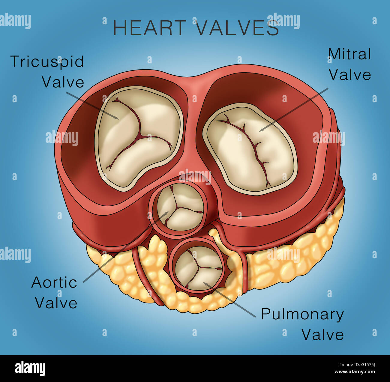
Pulmonary Tricuspid Aortic Mitral Valve Biological Stock Illustration Find pulmonary tricuspid aortic mitral valve biological stock images in hd and millions of other royalty free stock photos, 3d objects, illustrations and vectors in the shutterstock collection. thousands of new, high quality pictures added every day. Right atrioventricular valve. valva atrioventricularis dextra. 1 4. synonyms: tricuspid valve, valva tricuspidalis. understanding heart valves anatomy is important in grasping the overall function of the heart. the heart is one of the most important organs in the body. it is responsible for propelling blood to every organ system, including itself.

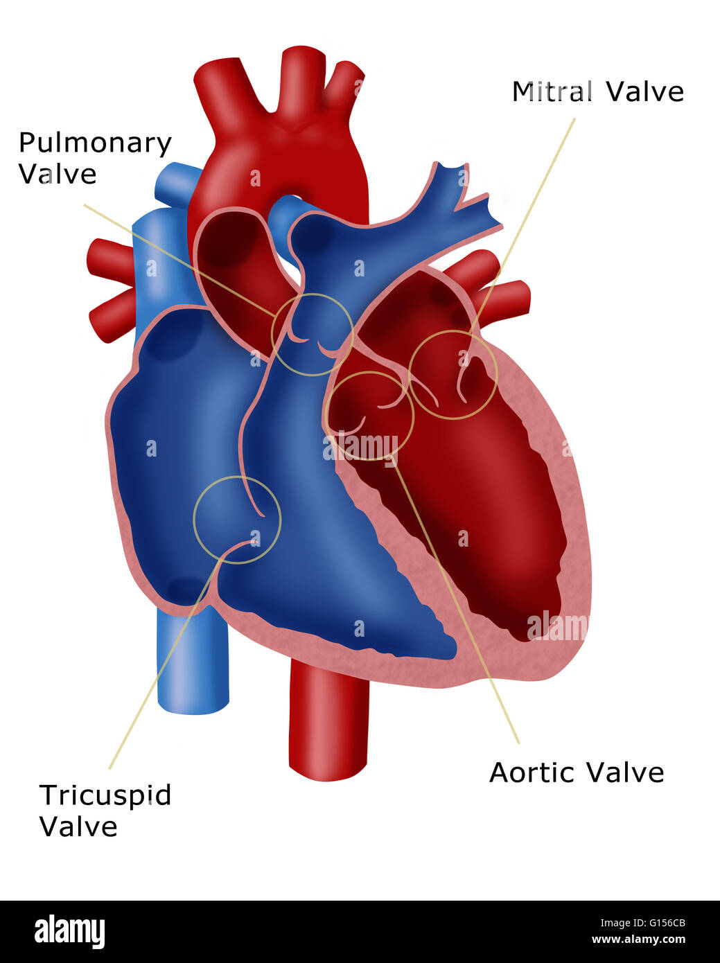
Illustration Of Heart Valves The Image Shown Includes The Mitral Valve Tricuspid valve disease is an often underrecognized clinical problem that is associated with significant morbidity and mortality. unfortunately, patients will often present late in their disease course with severe right sided heart failure, pulmonary hypertension, and life limiting symptoms that have few durable treatment options. traditionally, the only treatment for tricuspid valve disease. The mitral and tricuspid valves are supported by the attachment of fibrous cords (chordae tendineae) to the free edges of the valve cusps. the chordae tendineae are, in turn, attached to papillary muscles , located on the interior surface of the ventricles – these muscles contract during ventricular systole to prevent prolapse of the valve. Heart valves. as your heart pumps blood, four valves open and close to make sure blood flows in the correct direction. as they open and close, they make two sounds that create the sound of a heartbeat. the four valves are the aortic valve, mitral valve, pulmonary valve and tricuspid valve. a heart murmur is often the first sign of a heart valve. In a study including 50 normal tricuspid valves (18), 5 types of chordae were distinguished by their morphology and mode of insertion: fan shaped, rough zone, basal, free edge, and deep chordae. the last 2 types are unique to the tricuspid valve. the number of chordae varied from 17 to 36 with an average of 25 chordae.

Illustration Of A Heart Showing The Four Valves Pulmonary Valve Heart valves. as your heart pumps blood, four valves open and close to make sure blood flows in the correct direction. as they open and close, they make two sounds that create the sound of a heartbeat. the four valves are the aortic valve, mitral valve, pulmonary valve and tricuspid valve. a heart murmur is often the first sign of a heart valve. In a study including 50 normal tricuspid valves (18), 5 types of chordae were distinguished by their morphology and mode of insertion: fan shaped, rough zone, basal, free edge, and deep chordae. the last 2 types are unique to the tricuspid valve. the number of chordae varied from 17 to 36 with an average of 25 chordae. When the ant leaflet was in view (14 cases), the aortic valve was visible in all cases, and in 13 of 14 cases, all three leaflets of the aortic valve were also visualized. in this view, the ant leaflet was seen as a single leaflet (figure 5a, videos 1 and 2). when the a p combination was seen (43 cases), the aortic valve was always in view, and. Pulmonary, tricuspid, aortic and mitral valve. biological valves and mechanical valves 3d illustration human heart tricuspid and bicuspid valve for medical concept.

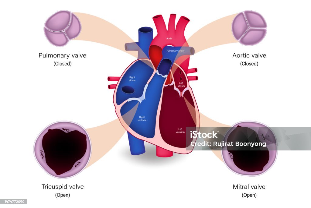
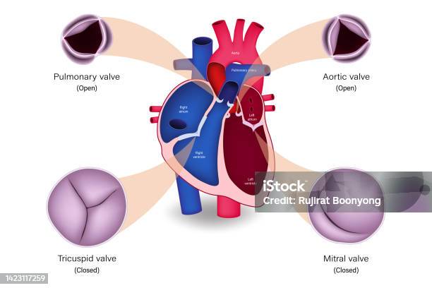
Human Heart Valve Anatomy Diastole Pulmonary Valve Aortic Valve When the ant leaflet was in view (14 cases), the aortic valve was visible in all cases, and in 13 of 14 cases, all three leaflets of the aortic valve were also visualized. in this view, the ant leaflet was seen as a single leaflet (figure 5a, videos 1 and 2). when the a p combination was seen (43 cases), the aortic valve was always in view, and. Pulmonary, tricuspid, aortic and mitral valve. biological valves and mechanical valves 3d illustration human heart tricuspid and bicuspid valve for medical concept.

Human Heart Valve Pulmonary Valve Aortic Valve Tricuspid Valve And

Human Heart Valve Anatomy Systole Pulmonary Valve Aortic Valve

Comments are closed.