Structure Of The Heart Valves Anatomy Mitral Valve Pulmonary

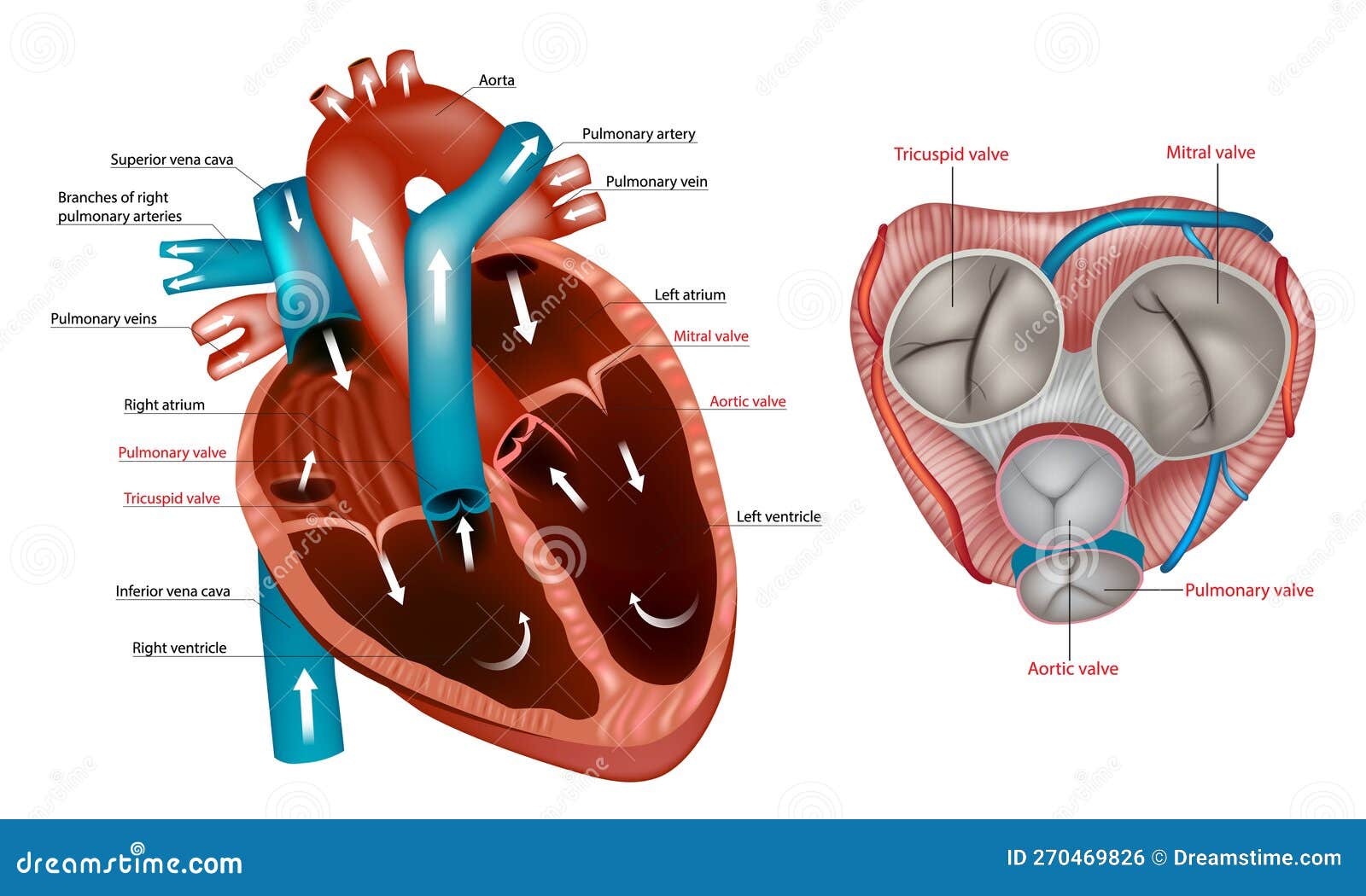
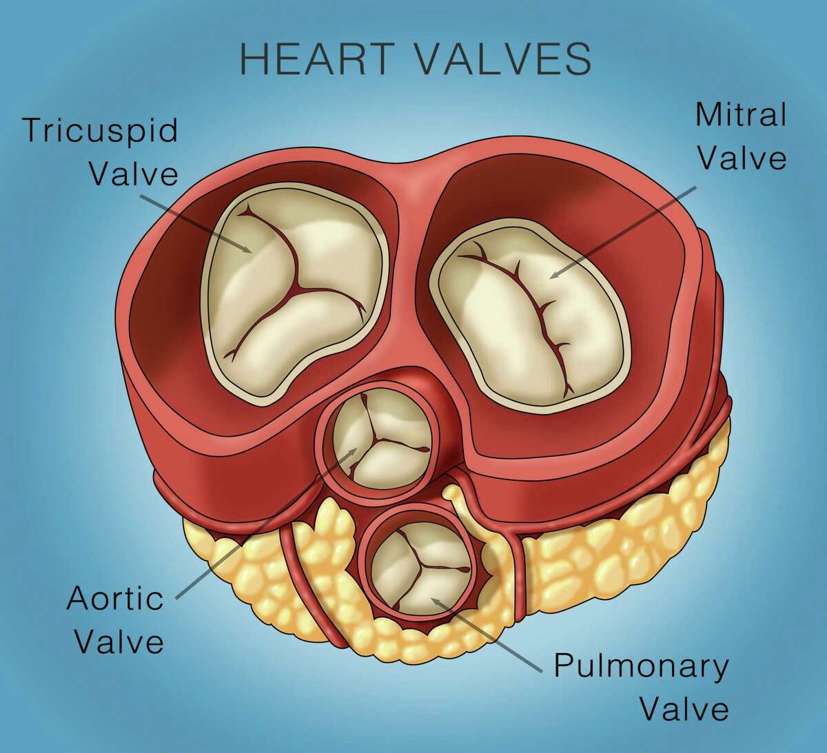
Structure Of The Heart Valves Anatomy Mitral Valve Pulmonary Valve Right atrioventricular valve. valva atrioventricularis dextra. 1 4. synonyms: tricuspid valve, valva tricuspidalis. understanding heart valves anatomy is important in grasping the overall function of the heart. the heart is one of the most important organs in the body. it is responsible for propelling blood to every organ system, including itself. They are composed of connective tissue and endocardium (the inner layer of the heart). there are four valves of the heart, which are divided into two categories: atrioventricular valves: the tricuspid valve and mitral (bicuspid) valve. they are located between the atria and corresponding ventricle. semilunar valves: the pulmonary valve and.

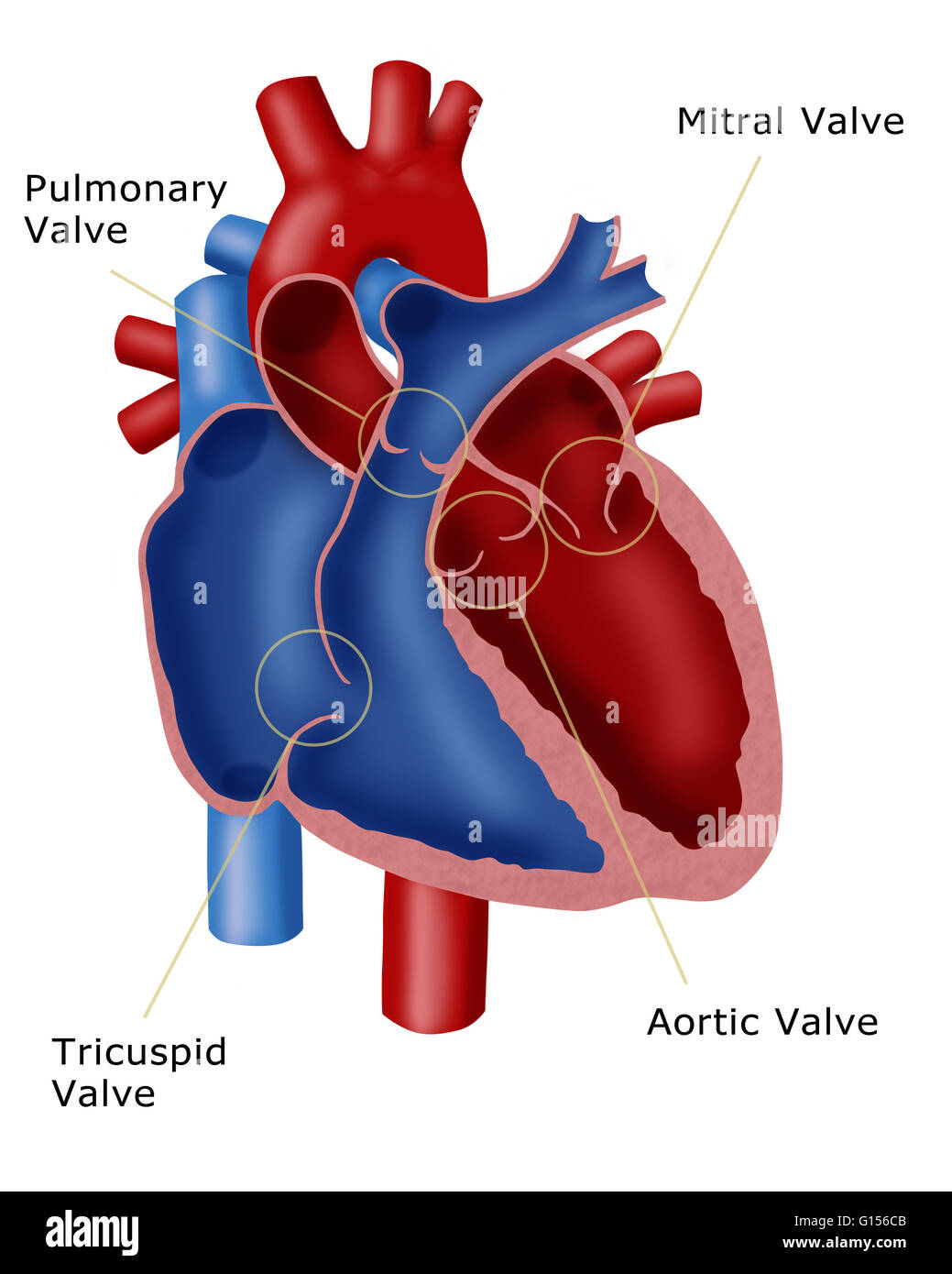
Heart Valve Anatomy Britannica Heart valves. as your heart pumps blood, four valves open and close to make sure blood flows in the correct direction. as they open and close, they make two sounds that create the sound of a heartbeat. the four valves are the aortic valve, mitral valve, pulmonary valve and tricuspid valve. a heart murmur is often the first sign of a heart valve. Mitral valve: located between the left atrium and left ventricle, the mitral valve ensures blood does not flow backward into the left atrium when the left ventricle contracts. semilunar valves : pulmonary valve : situated between the right ventricle and pulmonary trunk, this valve prevents the return of blood into the right ventricle after it. The heart has four chambers; the right and left atria and right and left ventricles. the heart also has valves that help with blood circulation, the mitral and tricuspid valves are called “atrioventricular” because of their location that is between the atriums and ventricles, and the aortic and pulmonary valves are the “arterioventricular” valves located between the ventricles and. The heart valves are especially important to effectively maintain the systolic and diastolic phase of the cardiac cycle. there are two types of heart valves; the atrioventricular valves (mitral, tricuspid) and the semilunar valves (aortic and pulmonic). the pulmonic valve physically separates the right ventricle from the pulmonary trunk. while.

Illustration Of A Heart Showing The Four Valves Pulmonary Valve The heart has four chambers; the right and left atria and right and left ventricles. the heart also has valves that help with blood circulation, the mitral and tricuspid valves are called “atrioventricular” because of their location that is between the atriums and ventricles, and the aortic and pulmonary valves are the “arterioventricular” valves located between the ventricles and. The heart valves are especially important to effectively maintain the systolic and diastolic phase of the cardiac cycle. there are two types of heart valves; the atrioventricular valves (mitral, tricuspid) and the semilunar valves (aortic and pulmonic). the pulmonic valve physically separates the right ventricle from the pulmonary trunk. while. The valves prevent the backward flow of blood. these valves are actual flaps that are located on each end of the two ventricles (lower chambers of the heart). they act as one way inlets of blood on one side of a ventricle and one way outlets of blood on the other side of a ventricle. each valve actually has three flaps, except the mitral valve. The aortic semilunar valve is between the left ventricle and the opening of the aorta. it has three semilunar cusps leaflets: left left coronary, right right coronary, and posterior non coronary. in clinical practice, the heart valves can be auscultated, usually by using a stethoscope.

Mitral Valve Repair Minimally Invasive Heart Surgery Vs Sternotomy The valves prevent the backward flow of blood. these valves are actual flaps that are located on each end of the two ventricles (lower chambers of the heart). they act as one way inlets of blood on one side of a ventricle and one way outlets of blood on the other side of a ventricle. each valve actually has three flaps, except the mitral valve. The aortic semilunar valve is between the left ventricle and the opening of the aorta. it has three semilunar cusps leaflets: left left coronary, right right coronary, and posterior non coronary. in clinical practice, the heart valves can be auscultated, usually by using a stethoscope.

Comments are closed.